Home

Pacifique Négligence Pedigree attenuation in ct scan persuader Éclat Amuser

PET/CT and Attenuation Correction Artifacts

a) Axial CT scan of the brain showing an area of low attenuation in the...  | Download Scientific Diagram
a) Axial CT scan of the brain showing an area of low attenuation in the... | Download Scientific Diagram

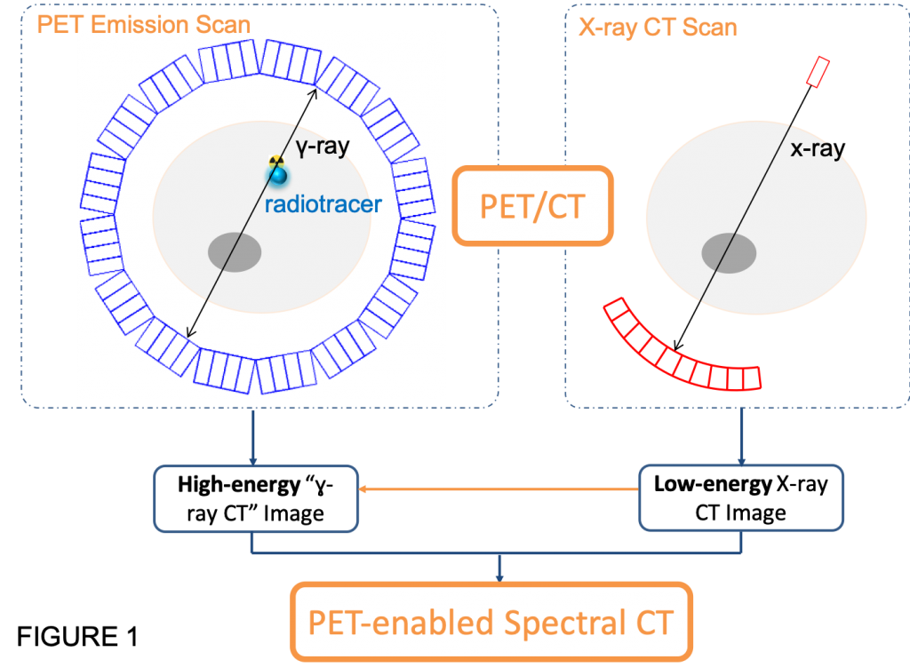
PET-enabled Spectral CT – Wang Lab – PET Parametric Imaging

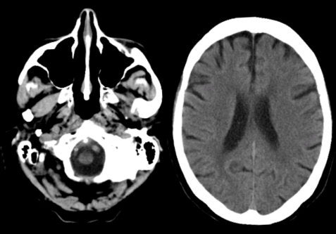
Common pitfalls of interpreting brain computed tomography (CT) scans |  Medmastery
Common pitfalls of interpreting brain computed tomography (CT) scans | Medmastery

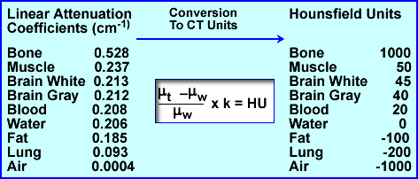
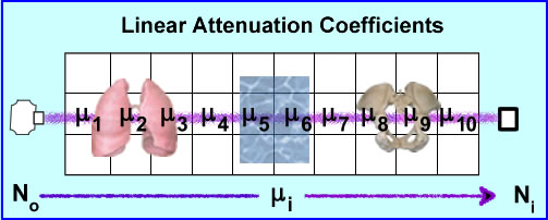
The steps involved in converting from a CT scan to linear attenuation... |  Download Scientific Diagram
The steps involved in converting from a CT scan to linear attenuation... | Download Scientific Diagram
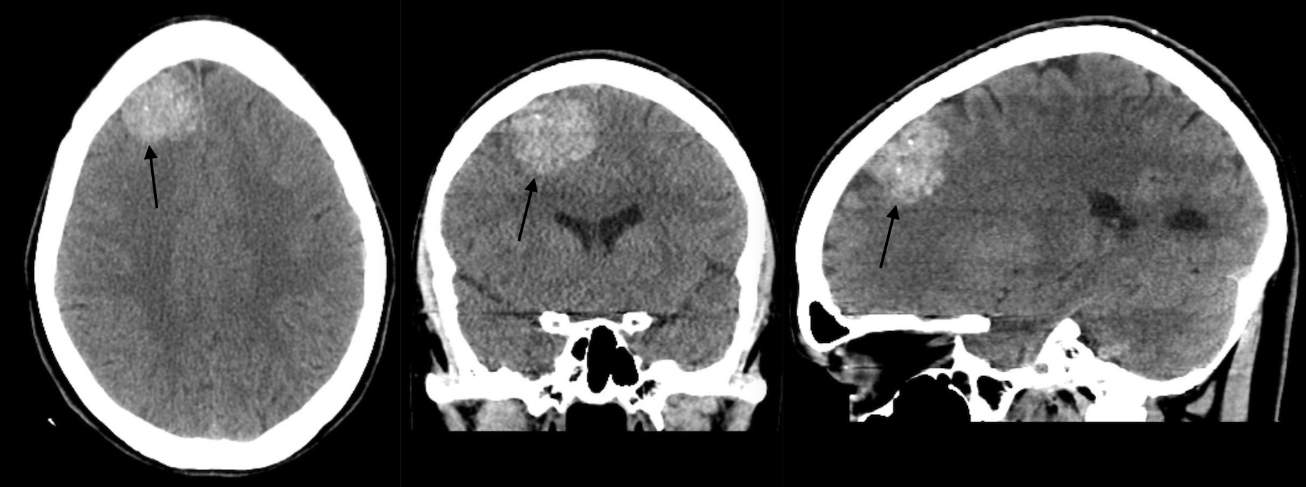
Fundamental Radiological Findings: Increased Signal Attenuation In The  Brain (Non-Contrast Head CT Scan) - Stepwards
Fundamental Radiological Findings: Increased Signal Attenuation In The Brain (Non-Contrast Head CT Scan) - Stepwards

Appearance of wooden foreign body on CT scan | Emergency Medicine Journal

Mosaic Attenuation: Etiology, Methods of Differentiation, and Pitfalls |  RadioGraphics
Mosaic Attenuation: Etiology, Methods of Differentiation, and Pitfalls | RadioGraphics

A, CT scan shows high-attenuation signals within the sulci of the... |  Download Scientific Diagram
A, CT scan shows high-attenuation signals within the sulci of the... | Download Scientific Diagram

Common pitfalls of interpreting brain computed tomography (CT) scans |  Medmastery
Common pitfalls of interpreting brain computed tomography (CT) scans | Medmastery

CT scans
CT scans

Common pitfalls of interpreting brain computed tomography (CT) scans |  Medmastery
Common pitfalls of interpreting brain computed tomography (CT) scans | Medmastery

CT Basics and PET Attenuation

CT Basics and PET Attenuation

A, CT scan shows slightly higher attenuation (white arrow) in the... |  Download Scientific Diagram
A, CT scan shows slightly higher attenuation (white arrow) in the... | Download Scientific Diagram

In non-contrast axial carnial CT scan; there is diffuse low attenuation...  | Download Scientific Diagram
In non-contrast axial carnial CT scan; there is diffuse low attenuation... | Download Scientific Diagram

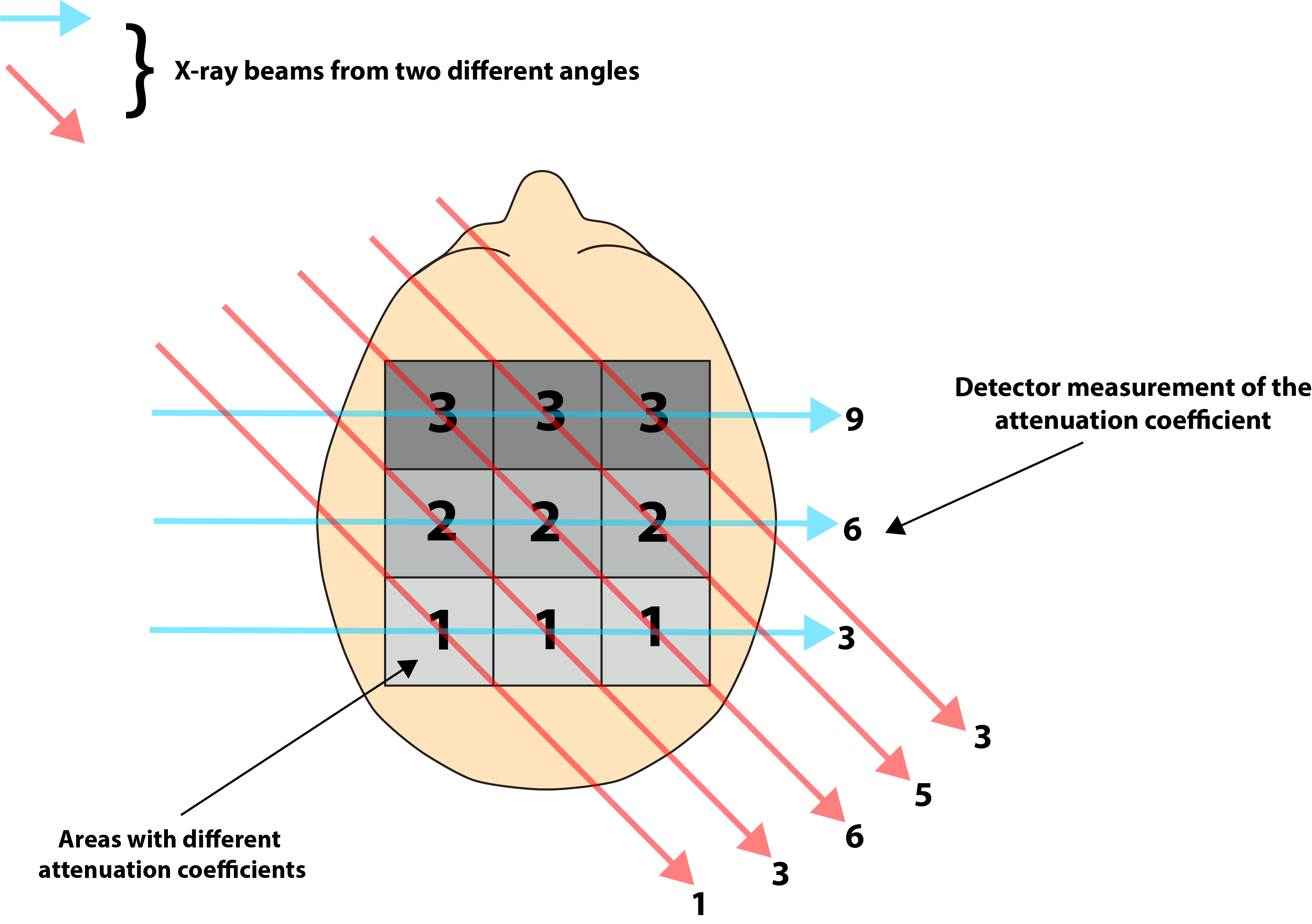
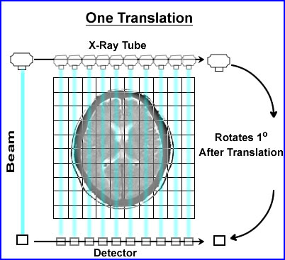
CT Basics
CT Basics

How to interpret an unenhanced CT Brain scan. Part 1: Basic principles of Computed  Tomography and relevant neuroanatomy
How to interpret an unenhanced CT Brain scan. Part 1: Basic principles of Computed Tomography and relevant neuroanatomy

Hemorrhage or not? Determining the cause of high attenuation on brain computed  tomography (CT) | Medmastery
Hemorrhage or not? Determining the cause of high attenuation on brain computed tomography (CT) | Medmastery

CT scan of the brain. A large low attenuation area in the territory of... |  Download Scientific Diagram
CT scan of the brain. A large low attenuation area in the territory of... | Download Scientific Diagram

CT Basics and PET Attenuation

Automated measurement of liver attenuation to identify moderate-to-severe  hepatic steatosis from chest CT scans - European Journal of Radiology
Automated measurement of liver attenuation to identify moderate-to-severe hepatic steatosis from chest CT scans - European Journal of Radiology

Common pitfalls of interpreting brain computed tomography (CT) scans |  Medmastery
Common pitfalls of interpreting brain computed tomography (CT) scans | Medmastery

Recognizing intra-axial tumors on brain computed tomography (CT) and  magnetic resonance imaging (MRI) | Medmastery
Recognizing intra-axial tumors on brain computed tomography (CT) and magnetic resonance imaging (MRI) | Medmastery